Difference Between Light and Electron Microscope (with Comparison Chart) |
您所在的位置:网站首页 › light microscope › Difference Between Light and Electron Microscope (with Comparison Chart) |
Difference Between Light and Electron Microscope (with Comparison Chart)
|
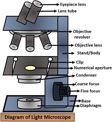
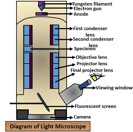
The difference between light and electron microscope is mainly due to the two properties like one is the source of illumination, and the second is the type of lens. Source of illumination: It is the property of a microscope that ensures the clear visibility of the object or specimen and adds brightness to it. Lens: It is used in the microscope, which can vary with different types of microscope available, and its primary function is to magnify the image. The light microscope uses a direct source of light waves to form the image, whereas the electron microscope uses a beam of an accelerated electron. In the light microscope, the glass-based lens is used, and it has a combination of eye-piece, objective and condenser lens. In the electron microscope, the electromagnetic lens is used, and it has a combination of condenser, objective and projector lens. Here, we will discuss the key differences along with the comparison chart between the light and electron microscope. Content: Light Vs Electron Microscope Comparison Chart What is a Microscope? Definition Diagram Key Differences Conclusion Comparison Chart PropertiesLight microscopeElectron microscope DiscoveryGiven by Zoocharia Janseen in 1590Given by Ernst Ruska and Max Knoll in 1931 Source of illuminationLight raysBeam of electron Lens usedEye-piece, objective and condenser lensCondenser, objective and projector lens Lens typeLens are of glass materialLens are of electromagnetic material Magnifying power1000X10,00,000X Resolving power0.2 µm0.001 µm Viewing screenImage is viewed directly through eye-pieceImage is viewed on fluorescent screen Power supplyRequires low power supplyRequires high power supply Cooling systemAbsentPresent Sample preparationSimpleComplex WorkingEasy to operateRequires technical skills to operate TypesMainly of four types: Bright field, Dark field, Phase contrast and Fluorescence microscopeMainly of three types: SEM, TEM and STEM Vacumn systemAbsentPresent CostCheapExpensive MagnificationLow, detailed structure cannot be studiedHigh, gives 3D structure of an object Specimen used5 µm thick specimen can be easily visualizedOnly thin specimen up to 0.1 µm can be visualized Image obtainedColouredBlack and white Filament usageAbsentTungsten filament is used as an electron source Radiation leakageAbsentPresent For contrast of the imageSpecimen is stained with dyesSpecimen is coated with heavy metals What is a Microscope?A microscope is a device, which comprises a set of lenses that allow us to see the magnified view of an object or a specimen. This device helps us to study the internal and external structures of the specimen that would not be possible without the use of a microscope. As from the name microscope, it is obvious that micro is a term used for minute things, and scope is a term used to look out things. Therefore, the microscope is an instrument that allows us to look or to see the microorganisms that are invisible to the naked eye. The study of the organisms under the microscope is called Microscopy. Definition of Light MicroscopeIt is also called a compound microscope. A light microscope is an optical microscope, which uses a ray of light to view the image where a condenser collects the light and diverges it to the specimen. It has comparatively a low-resolution and magnification power than the electron microscope, which is about 0.2 µm and 500 – 1000 X, respectively. Definition of Electron MicroscopeIt is an optical microscope, which uses a beam of an accelerated electron from the source of heated tungsten filament that transmits the electron to the specimen. It results in a high-resolution image of 0.001 µm resolving power, i.e. 250 times more than the light microscope and high magnifying power of 10,00,000 X. Diagram of a Light MicroscopeIt consists of several components like:
It has several elements which are as follows:
So finally, we have discussed the difference in properties, structure and components of both the light and electron microscope. Therefore, we can conclude that both the light and e– microscope works on the different principles, in which one uses a light source, and the other uses the electron to form the image. The construction and operation of both the microscopes are different, where a light microscope is easy to handle, and the electron microscope requires technical skills to operate. You Might Also Like:AIDsThymus Dependent AntigensSpectrophotometerChloroplast in Plant CellFluorescence Microscopy |
【本文地址】